
Summary
Scientific article on veterinary radiographic projections and contrast media. Positioning techniques, routine and oblique projections, positive and negative contrast agents, and their importance in diagnostic imaging for domestic animals are explained.
What You Will Learn
- Principles for naming radiographic projections.
- Differences between routine and oblique projections.
- Clinical importance of positioning.
- Types of contrast media and their use.
- The role of the illuminator (negatoscope) in diagnostic evaluation.
- Technical impact on the accuracy of veterinary diagnosis.
Introduction
The diagnostic quality of an X-ray does not depend exclusively on the equipment used, but on the correct positioning of the patient and the appropriate selection of the radiographic projection. Inadequate technique can lead to superimpositions, anatomical distortions, or erroneous interpretations.
In veterinary medicine, where patient cooperation is limited and anatomical variations between species are significant, technical precision acquires special relevance. This article develops the foundations of radiographic projections, their classification, clinical applications, and the use of contrast media as complementary tools in diagnostic imaging.
1. Radiographic Projections: Concept and Foundation
Radiographic projections are named according to the direction the central ray of the primary beam follows from the point of entry to the point of exit in the animal's body.
The goal is to obtain the most convenient posture for the patient, reduce unnecessary superimpositions, and provide an image with diagnostic value. The correct choice of projection allows for more precise visualization of specific anatomical structures.
2. Routine Radiographic Projections
Routine projections constitute the basis of radiological studies in veterinary medicine. The main ones include:
- 2.1 Ventro-Dorsal (VD): The ray enters through the ventral surface and exits through the dorsal. Common in abdominal and thoracic studies.
- 2.2 Dorso-Ventral (DV): The ray enters through the dorsal region and exits through the ventral. Indicated when the VD position is not clinically viable.
- 2.3 Cranio-Caudal (Cra-Cad): Used mainly in limbs, allowing for the evaluation of bone alignment.
- 2.4 Caudo-Cranial (Cad-Cra): The inverse of the above, useful depending on the anatomical structure being evaluated.
- 2.5 Right Lateral (RL) and Left Lateral (LL): These allow for the evaluation of depth and three-dimensional anatomical relationships in the thorax and abdomen. Bilateral comparison is fundamental in orthopedic studies.
3. Special Projections in Limbs
In distal structures such as metacarpals and metatarsals, especially in equines, the following are used: Dorso-Palmar (DP), Palmo-Dorsal (PD), Dorso-Plantar (D-Pl), and Planto-Dorsal (Pl-D). These projections allow for the evaluation of cortical integrity, joint spaces, and the presence of osteolytic or proliferative lesions.
4. Oblique Projections
Oblique projections are employed when standard projections do not allow for adequate visualization due to bone superimposition. Examples include:
- Dorso 60° Lateral-Palmar Medial Oblique (D60°L-PaMO).
- Dorso Medial-Palmar Lateral Oblicua (DM-PaLO).
- Palmar Medial-Dorso Lateral Oblicua (PaM-DLO).
- Palmar Lateral-Dorso Medial Oblicua (PaL-DMO).
These projections are especially relevant in equine orthopedic diagnosis, where minor alterations can affect athletic performance.
5. Importance of Correct Positioning
Inadequate positioning can generate geometric distortion, false asymmetries, superimposition of structures, and diagnostic errors. In veterinary radiology, proper restraint and sedation when necessary are essential to ensure accuracy.
6. Radiological Contrast Media
Contrast media are chemical substances used to modify the radiographic density of structures that are normally not visible. Their purpose is to delimit cavities, evaluate hollow organs, and determine the size, shape, and position of soft tissues. They can be administered orally or intravenously.
7. Types of Contrast Media
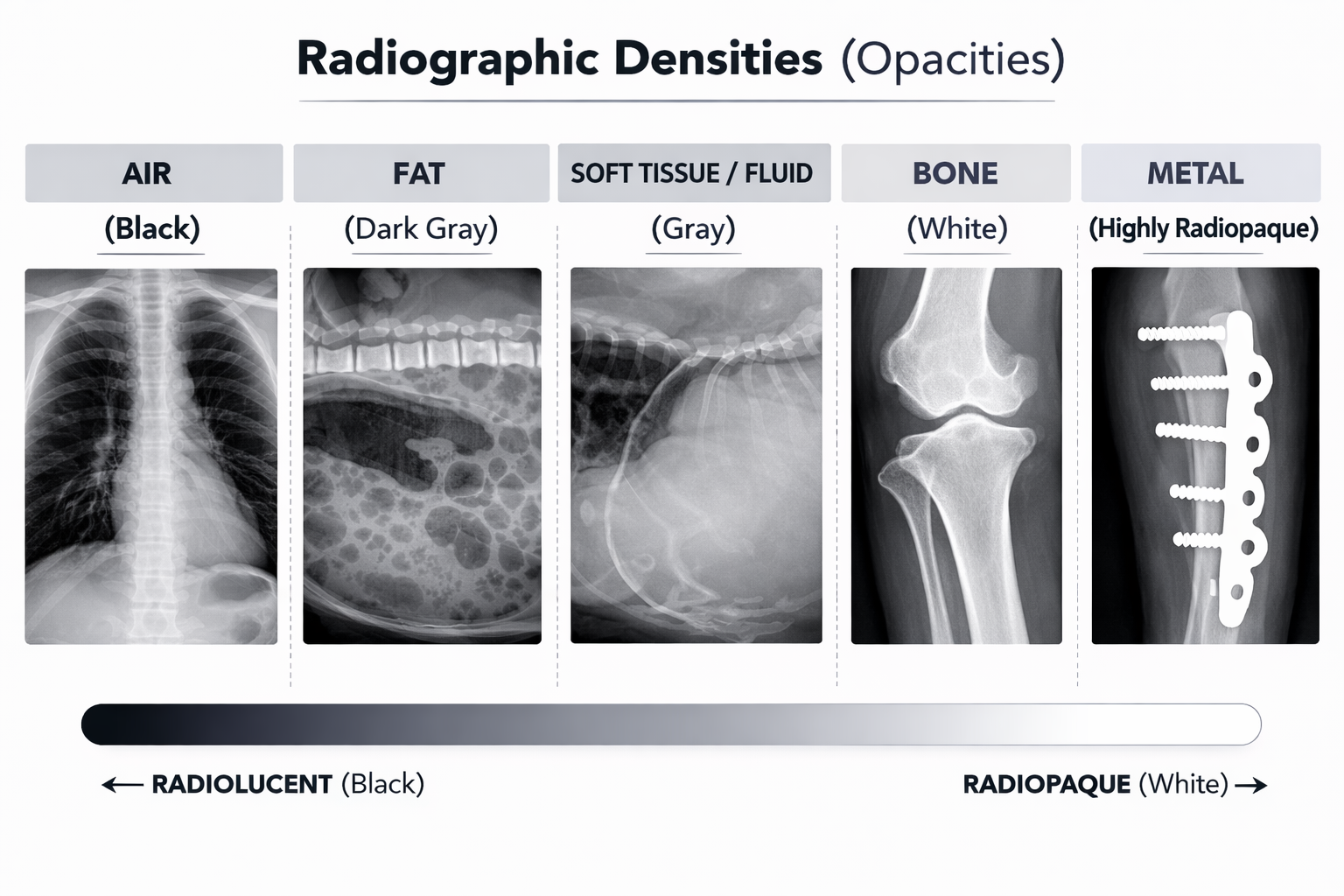
7.1 Positive Contrast Agents
They contain elements with a high atomic number, making them radiopaque. Example: Barium sulfate. They appear white on the X-ray and allow for visualization of the gastrointestinal tract.
7.2 Negative Contrast Agents
They are low-density gases, making them radiolucent. Examples: Air, Oxygen, Carbon dioxide. They are used to create internal contrast and delimit cavities.
8. The Illuminator (Negatoscope) in Radiographic Evaluation
The negatoscope is the device used to examine radiographic plates. Uniform lighting is essential to correctly evaluate densities and identify subtle details. In modern digital systems, this function is performed by calibrated monitors.
9. Clinical Application and Diagnostic Relevance
The correct selection of projection and contrast allows for the diagnosis of hidden fractures, identification of joint displacements, evaluation of pulmonary lesions, and detection of gastrointestinal obstructions. Technical precision is decisive in avoiding incorrect treatments.
Conclusion
Radiographic projections are an essential component of diagnostic imaging. Their correct execution minimizes superimpositions and optimizes anatomical visualization. The use of contrast media expands diagnostic capacity by evaluating structures not visible in simple studies. Technical mastery of these tools is fundamental in modern veterinary practice.